

Hand-raising Orphan Asian Elephants
Chapter 9: Medical Procedures
By Susan K. Mikota
With contributions from Bhaskar Choudhury, Vijitha Perera, and Willem Schaftenaar
Sections in this chapter include the following:
-
Introduction
-
Anesthesia and sedation
-
Basic supplies and equipment
-
Bandages, braces, casts, and splints
-
Blood collection
-
Blood transfusion
-
Breast milk collection
-
Culture collection – Blood
-
Culture collection – Wounds
-
Enema
-
Fecal analysis
-
Fecal transplantation
-
Fluid therapy - IV / rectal
-
Injections – IM, SQ, IV
-
Oxygen therapy
-
Postmortem exam
-
Radiographs
-
Urine collection and analysis
-
Vaccinations
-
Wound treatment techniques
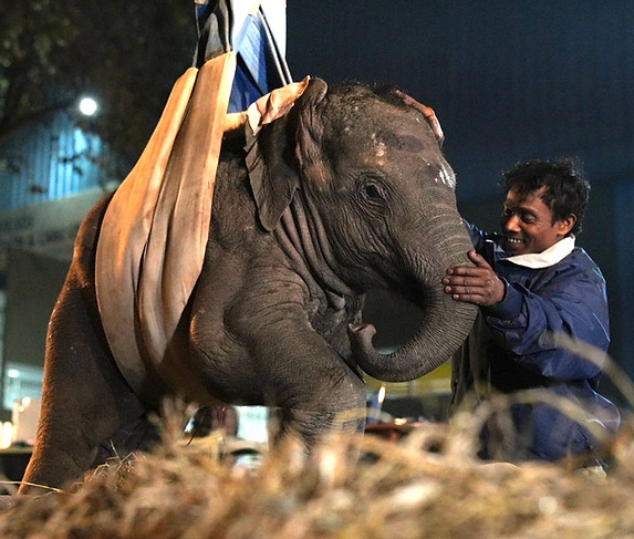
Photo credit Wildlife SOS, India.
Introduction
This chapter describes some of the medical procedures that may be needed to care for orphan elephants. Any medical interventions should be conducted by or under the supervision of a veterinarian.
Anesthesia and Sedation
Anesthesia/sedation may be required for transport or to conduct diagnostic or treatment procedures. The risk of using an anesthetic/sedative agent in a young elephant is usually acceptable as long as good practices are followed. Many procedures can be done under standing sedation See also: Anesthesia | Elephant Medicine.
Calves a year of age or more (height at shoulder of 120-150 cm) can be difficult to restrain physically at the rescue location and may be quite aggressive and capable of causing serious damage to handlers. Sedation is a safe option to protect both elephant and human.
Sedation helps in conducting a detailed clinical examination without stressing out the animal, which otherwise requires forceful restraining. All rescued calves invariably are moderately to severely dehydrated, and sedation facilitates correcting the fluid balance at the rescue site using IV injections at multiple sites which otherwise is difficult in a physically-restrained calf.
Transportation of orphan calves to a rehabilitation facility can also be quite stressful. The stress of transport may result in food refusal and a struggle to adapt to the the new environment at the rehabilitation facility. Much of this stress can be avoided by using moderate sedation during transport. Xylazine (0.08 mg/kg IM) together with ketamine (0.10 mg/kg) has been used for sedation and transportation of very young calves without any complications for distances ranging from 100 to 350 kilometers (Choudhury, B and Talukdar, A unpublished). The calves were loaded into the transport trucks and placed in lateral recumbency (on their sides) before injecting the anesthetic agents. The calves were accompanied by at least two handlers. Supplemental ketamine was given IM in few cases when necessary.
Xylazine can be used to induce mild to deep sedation with stable physiologic variables and no major side effects. For mild sedation, Dr. Perera recommends 20 mg IM for the first 100 kg of body weight plus 10 mg for every additional 100 kg. For deep sedation, give 40 mg xylazine IM plus 20 mg for every additional 100 kg. Supplemental doses (generally at half of the original dose) can be given if needed.
The response to xylazine sedation may vary between individuals. Loud noises can stimulate arousal so sedation should take place in a quiet environment. Xylazine sedation can be reversed using yohimbine at 1 mg yohimbine per 10 mg xylazine (0.05-0.13 mg/kg IV). Atipamizole is also an effective reversal agent but may not be as widely available and is more expensive. Give 1 mg of atipamizole IM or slow IV for every 10 mg of xylazine.
Note: It is a good idea to cover the eyes of a sedated calf to help maintain a calm state and prevent external visual stimulation.
Once at the rehabilitation facility, the presence of adult captive elephants can help older calves stabilize in their new environment. Calves older than a year or more who are not trained to avoid solar fences can struggle in a new environment and become unmanageable. Tying a jute rope around the belly of the calf and attaching it to and adult elephant initially may help. This should be done under observation. If the calf struggles, even soft ropes can cause rope burns.
Basic Supplies and Equipment
Following is a comprehensive list of equipment and supplies that may be needed to carry out the medical procedures that are discussed in this chapter. Larger facilities dedicated to elephant/wildlife rescue and with veterinarians on staff will likely have more of these items on site; small facilities may only need to stock basic items (indicated with a *) and use nearby veterinary clinics or universities for special needs. See Tables 9.1, 9.2, and 9.3.


In addition to the above items, oral, injectable, and topical medications may be stocked as needed. Available products may vary between countries. Commonly used antibiotics for calves include ampicillin (injectable only), amoxicillin, penicillin G procaine (long-acting), and ceftiofur. For severe infections amikacin or gentamicin may be indicated (Emanuelson, 2006).
Ideally antibiotic choice should be guided by culture and sensitivity results (antibiogram). Maintain hydration when giving antibiotics, especially when using aminoglycosides such as amikacin or gentamicin. Tetracyclines and fluoroquinolones are best avoided in growing animals. Ceftiofur long‐acting (Excede) 6.6mg/kg SC gives effective plasma levels for 7–10 days. Pharmacokinetic studies have not been conducted in young elephants so refer to studies that have been done in adult elephants. Refer to the ECI Formulary for more information.
Bandages, Braces, Casts and Splints
Bandages, braces, casts, or splints should only be applied by veterinarians or trained personnel. They should be monitored daily by care staff to make sure they stay dry and are not too tight. Improperly applied external devices can interfere with circulation. Any external devices that become wet or soiled should be changed. See Figures 9.1, 9.2, and 9.3.
Figure 9.1 shows a bamboo splint and cloth bandage on the foot of a four-month-old elephant rescued from a hunting trap in a forest in southwestern Myanmar, and being cared for at the Wengabao Elephant Sanctuary northeast of Yangon. Read the full article here.
A Robert Jones bandage and then a fiberglass cast were used to treat an ulna fracture in a two-year old elephant (Karunarathne et al., 2017). See Figure 9.2. Read the full article here.

Figure 9.2 Young elephant with a cast.

Figure 9.1 A bamboo splint and cloth bandage on four-month-old calf.

Figure 9.3 Splint applied to stabilize front leg fracture. Photo credit Bhaskar Choudhury.
Blood Collection
The ear (auricular) and secondly the hind leg (saphenous) are the most commonly used veins for blood collection. The ear veins may be accessed with the elephant standing, however, the veins are more prominent with the elephant in lateral recumbency (laying on the side and using the down ear). In this position gravity helps the veins to fill. It is also the safest position for the veterinarian should the elephant move suddenly. The ear veins are most visible on the back (caudal aspect) of the ear but can be seen on the front (rostral) aspect of the ear in some elephants. See Figure 9.4.
The ear veins are thinner walled than adjacent arteries. Arteries are turgid and a pulse can be palpated. Veins have thin walls and collapse when pressed.
A pulse can generally be detected in arteries for further differentiation. When the environmental temperature is low, the ear veins may collapse; flushing the ear with warm water for several minutes may help to increase the blood flow locally.
The hind leg (saphenous) vein is on the inside (medial aspect of the leg). It can be accessed most safely with the elephant in lateral recumbency. The saphenous vein is deeper than it looks.
The front leg vein (cephalic) is another site that can be accessed with the elephant standing but presents a risk to the operator should the the head or trunk move suddenly. See also: Hematology | Elephant Medicine.
Blood can be collected using a syringe and needle, a vacutainer and needle or a winged blood collection set. There are a number of disadvantages to using a syringe and needle. If the elephant moves the needle may come out, the vein may be traumatized and you will have to stick the elephant again. If an inadequate amount of blood is placed in the purple top tube, the hematocrit may be artificially reduced due to dilution with the anti-coagulant in the tube (EDTA).

Figure 9.4 Ear vessels in a young elephant. Arteries are turgid and a pulse can be palpated. Veins have thin walls and, when pressed, the proximal part of the vein (between the pressing finger and the head) becomes more visible as it will swell when it fills with stagnating blood. Photo credit: Willem Schaftenaar
Winged blood collection sets are convenient for collecting blood and offer several advantages. The set consists of a winged needle for placement in the vein connected by tubing to a needle that punctures the tube and a plastic holder. The plastic holder helps to stabilize the tubes. See Figure 9.5 and inset image.


Figure 9.5 Blood collection in an adult elephant using a winged blood collection kit. Inset: image of collection device. Photo credit: Willem Schaftenaar
Blood collection sets allow the operator to move with the elephant without dislodging the needle from the vein. Blood can be collected directly into vacutainer tubes so multiple tubes can be collected. The vacuum draws sufficient blood for the proper anticoagulant ratio in tubes such as EDTA. Collection directly into a vacuum tube reduces platelet clumping and clot formation when collecting samples for CBCs. Even small clots will reduce the platelet count and the hematocrit and WBC count may be affected if there are a lot of clots. Bleeding kits with 21 ga needles can be used for calves.
In very small calves or in situations such as shock where blood pressure is low, the draw from a vacutainer tube may collapse the vein. In these situations, a needle and syringe may be preferable, drawing slowly.
Blood is typically collected into a purple/lavender top tube (EDTA) for a complete blood count (CBC) and a red top tube for serum chemistries. Proper technique is important to prevent hemolysis (rupture of red blood cells) which can alter several lab results. To prevent hemolysis:
-
Swab the vein with alcohol but allow it to dry before inserting the needle.
-
Use the largest needle that will work.
-
If the syringe or tube is filling slowly, gently adjust the needle - the tip may be pressed against the vein.
-
If you are transferring the blood from a syringe to the tube allow the blood to flow down the side of tube so there is no splashing.
-
Don’t force the blood into the tube– allow the suction from the tube to draw the blood in. Don’t shake the tube. When using a tube without vacuum, remove the needle from the syringe before transferring the blood to the tube.
-
Purple/lavender top tubes contain an anticoagulant (EDTA). The tube should be gently inverted it so that the EDTA mixes with the blood sample.
-
Don’t expose your samples to extreme temperatures. Store tubes in a cold box or the refrigerator until they can be transferred to the lab.
Blood Transfusion
Indications for blood transfusion include blood loss from trauma or surgery or severe anemia (which can be due to multiple causes). As in other species, it appears that there are different blood groups in elephants and incompatibility may occur (Bansiddhi et al., 2015).
Blood compatibility testing (cross-matching) should be performed to determine if there are antibodies in the recipient’s blood that will react with the red blood cells of the donor. A cross-matching procedure in elephants is discussed in the following article: Blood compatibility testing in Asian elephants using an indirect antiglobulin technique to improve captive breeding success. Read full article here.
A cross-matching procedure can also be found in the Guidelines for Management of Elephant Endothelial Herpesvirus (EEHV) in Asia. (See also: Elephant Endotheliotropic Herpes Virus 1 | Elephant Medicine).
Blood Transfusion in Equids—A Practical Approach and Review (Jamieson et al., 2022) is an excellent review of blood transfusion in the horse and discusses many aspects that are also applicable to elephants. It includes a formula for calculating the blood volume needed by a recipient and how much can be collected from a donor, a description of the blood collection bags that must be used and practical tips to perform the procedure. Read the full article here.
Breast Milk Collection
Passive immunity is the transfer of antibodies to a newborn animal either prenatally (before birth) via the placenta, or postnatally (after birth) via ingestion of colostrum. Colostrum is the the first milk secreted after birth. It is rich in immunoglobulins (antibodies) that help to prevent disease in the first few weeks of life while the neonate’s own immune system is developing. In domestic species that depend on postnatal antibody transfer (cows and horses for example), newborns that do not receive colostrum are more likely to experience medical problems and have higher mortality. This is called failure of passive transfer.
Two studies have demonstrated that maternal to fetal antibody transfer does occur prenatally via the placenta in elephants (Nofs et al., 2013; McGee et al., 2014).
Our knowledge of the elephant’s immune system is not complete and while it has been shown that prenatal antibody transfer occurs in elephants, we don’t know if colostrum also plays a role.
In captive situations where the mother cow is present but the newborn elephant calf fails to nurse, attempts to collect colostrum can be done by hand or by using a human breast pump. However, milking manually is extremely difficult in most adult elephants. Some cows do not cooperate at all and even those that do cooperate may only yield a small volume. Giving oxytocin (~2 commercial units) to the cow 5 minutes prior can facilitate milk let-down.
To collect milk manually, squeeze the teat at the top with the thumb and forefinger and then squeeze the other three finger in succession. The procedure is similar to milking a goat.
If the cow will tolerate it, a breast pump can be applied for 10-20 minutes to each breast. Applying warm water packs, massaging, and allowing rest periods may help to increase the volume (Emanuelson, 2006).
Note: Remember that that the passage of antibodies colostrum can only occur for a few days after the birth so the procedure of collecting milk as described above is not likely applicable to most orphan situations. See comment on gut pathogen protection in Chapter 4.
Culture - Blood
A blood culture is performed in critically ill animals to determine whether infection-causing microorganisms are present in the bloodstream and causing sepsis (septicemia). Sepsis is discussed in Chapter 8. Special collection bottles – one for aerobic and one for anaerobic organisms are needed. A detailed procedure must be used to collect the sample to avoid contamination. If a blood culture is indicated, contact the lab where you will be sending the samples to obtain the correct bottles and follow the collection procedure they recommend.
Culture - Wound
Not all wounds need to be cultured but cultures are essential if you are dealing with an infected or non-healing wound. Use aseptic technique. Wear sterile gloves. Clean the tissue around the wound with a scrub to avoid contaminating the sample with organisms from the surrounding skin. Avoid getting the scrub in the wound. If there is pus or slough, irrigate the surface with sterile isotonic fluid to remove the more superficial material. When you insert your culture swab, avoid contacting the skin margins.
Collect samples from the deepest areas of the wound and sample multiple sites if there are pockets, fissures, or layers. If you suspect a biofilm, draw the swab across the surface of the wound with enough pressure to collect a sample of the biofilm but avoid causing any bleeding. In most wounds, bacteria are free-floating and act as individual organisms. But in chronic wounds, microbes attach and form invisible multicellular units called biofilms. Biofilms impair healing and they can be relatively impervious to common antiseptics, alcohols, bleach, and hydrogen peroxide.
When possible, include tissue samples from the wound. For example, debride the wound and submit some of the debrided material. Tissue samples are more likely to yield reliable culture results. Also submit any other material removed from the wound, such as a foreign body. Use a separate swab for each sample type – exudate, tissue, biofilm.
It’s a good idea to consult your lab for any special instructions on collection, transport, or shipping. When you submit samples, provide clinical information. Request cytology, aerobic and anaerobic culture, and antibiotic sensitivity testing. If available, request quantitative sensitives that provide Minimum Inhibitory Concentrations (MICs) for the drugs tested. If you suspect a fungal pathogen, request fungal cytology and culture. Collect all samples before starting or changing antibiotic therapy. If the elephant is already receiving antibiotic therapy, either suspend treatment, or collect the samples before the next scheduled dose. Ideally, stop treatment for a period that is at least 8 hours longer than the dosing interval. For example, for an antibiotic administered every 12 hours, stop treatment for at least 20 hours before collecting samples for culture. This interval allows the residual bacteria to re‐enter the log phase of active growth.
Enema
Enemas are used in cases of constipation or to administer fluids. If the calf is large enough to allow insertion of the hand, some feces can be removed manually. It is important to use lots of lubrication (e.g. mineral oil) on the tube to prevent irritation to the rectal mucosa. Warm water is best and can be administered via flexible tubing attached to a human enema bag, large syringe, or a funnel. Figure 9.7. The end of the tubing should be smooth (holding the end of the tubing in a flame will soften the edges). See also fluid therapy below.
When giving the enema because of constipation, gently move the tubing back and forth to break up the fecal boluses. The volume of fluid to use depends on the size of the calf and severity of the constipation.
Figure 9.7 Tubing and funnel to administer an enema. Photo credit: Susan Mikota.

Fecal Analysis
Feces from orphan elephant calves should be examined for the presence of intestinal parasites eggs. Parasites are discussed in Chapter 8. Collect a sample (~ 5 grams) from a fresh defecation. Store in a closed container in a cool box or refrigerator until the sample can be transported to the lab which should be as soon as possible. Commonly used flotation techniques are adequate to detect nematode eggs from fresh samples. An alternative method assesses nematode larvae and may be better for older samples (Abeysinghe et al., 2012).
Lynsdal et al. investigated the the effects of different collection and storage methods on helminth egg counts. They found that a fresh sample(s) collected within 7.5 hours of defecation was representative of the parasite load but
storage in formol saline and formalin significantly reduced the number of eggs recovered. However, fresh samples can be stored in anaerobic conditions (e.g. in sealed, zip-locked bags) at 4-6 °C for approximately 7 days without significant declines in egg recovery (Lyndsdale et. Al., 2015). They recommend a modified McMaster technique.
Trematode eggs are heavy and a sedimentation technique (such as the Baermann technique) may be needed. See Figures 9.8, 9.9, and 9.10 for examples of parasite eggs and larvae.

Figure 9.8 Examples of parasite eggs that may be seen under the microscope. A. Strongyle B Trematode; C. Ascarid; D. Strongyle (free-living larval form). From: Phuphisut O, Maipanich W, Pubampen S, Yindee M, Kosoltanapiwat N, Nuamtanong S, Ponlawat A, Adisakwattana P. Molecular identification of the strongyloid nematode Oesophagostomum aculeatum in the Asian wild elephant Elephas maximus. J Helminthol. 2016 Jul;90(4):434-40. (Phupisut et al., 2016).
%20ova.jpg)


Figure 9.9 Cestode (tapeworm), ova B. Strongyle, ova C. Larvated strongyle. Photo credits M. Ashokkumar, Centre for Wildlife Studies, Kerala Vet. and Animal Sciences University, Pookode, Wayanad. Abhijith TV, Ashokkumar M, Dencin RT, George C. Gastrointestinal parasites of Asian elephants (Elephas maximus L. 1798) in south Wayanad forest division, Kerala, India. J Parasit Dis. 2018 Sep;42(3):382-390.(Abhijith et al., 2018).
Fecal egg counts using techniques such as the McMasters count the number of eggs per gram of feces and provide an indication of the parasite load. Checking the egg count before and after anthelmintic treatment determines response to treatment.
At the Elephant Transit Home in Sri Lanka, if the eggs per gram count of nematodes is high calves are dewormed using fenbendazole, albendazole, levamisole, or ivermectin. If there are Fasciola eggs in the feces, they are treated with triclabendazole and if Anoplocephala eggs are found they are treated with praziquantel.
Fecal Transplantation (Transfaunation, Fecal Microbial Transplant)
Fecal transplantation is the process of transferring fecal bacteria from a healthy donor to a sick animal. It has been used in humans, cows, and horses. It is probably most useful for a calf with diarrhea although it was used in one adult elephant following an impaction (Greene et. al., 2019). The donor elephant should be healthy and ideally tested to verify that she is salmonella-free. If you are thinking of using this procedure, please refer to the article by Mullen (Mullen et al., 2018) for instructions and precautions. Read the article here.

Figure 9.10 Typical Fasciola ova. Source: https://www.ncvetp.org/uploads/7/4/9/2/74925531/6040603-orig_orig.jpg
Fluid Therapy
The daily maintenance fluid requirement for adult elephants (based on values for horses) is 40-60 ml/kg/24 hours and may be higher for calves. If the calf is not drinking at all, maintenance fluids must be supplied as well as additional fluids to replace any losses. This can be calculated as follows: replacement = body weight (kg) X % dehydration.
Rectal fluid administration is the preferred route in calves. Rectal administration is easier and quick acting compared to IV administration. Luke-warm water can be given 3 or 4 times daily up to every 2 hours in very sick calves at 10-20 ml/kg of body weight. If repeated administration of rectal fluids is needed, the anus of the elephant may become sensitive to touch. This can be addressed by mixing 15 ml of 2% lidocaine with some lubricant and applying it topically to the anus. Wait for 10 minutes before placing the tube in the rectum. After finishing the enema, the tail should be pressed down for a minute to prevent the loss of fluids.

Figure 9.11 Young calf receiving IV fluids. Photo credit: Vijitha Perera
IV fluid therapy can be considered if rectal therapy is not effective but it is challenging. If an IV catheter is placed, the calf will need to be monitored constantly to prevent removal. The ear or medial saphenous veins can be used. See Figure 9.11.
Note that the ear veins are susceptible to damage (necrosis) especially if medications are injected. A procedure for placing an IV catheter in a calf can be found in the Guidelines for Management of Elephant Endothelial Herpesvirus (EEHV) in Asia.
One option is to use intermittent bolus therapy, administering 20-40 ml/kg slowly over 60 minutes or more through an IV or butterfly catheter. If IV fluid is essential and the calf struggles or is very stressed, sedation may be required.
Example - How much fluid is needed?
A 200 kg calf has severe diarrhea and is not drinking. The calf is depressed, the mucous membranes are dry, and the capillary refill time is slow. The estimated dehydration is 5%. How much fluid do you need to give?
1. Calculate the maintenance needs: 40 ml/kg X 200 kg = 8000 ml (8 liters)/24 h
2. Calculate the replacement fluids needed (to replace the fluids lost due to diarrhea):
-
Body weight X % dehydration = volume to replace
-
200 kg X 5% = 1000 ml = 1.0 liters /24 h
The total amount of fluid to be given over 24 h is 8 liters + 1.0 liters = 9.0 liters.
Fluids can be given by one route (e.g. rectal) or by using multiple routes (rectal + oral; rectal + IV; or rectal + oral + IV). Elephant blood is hypo-osmotic compared to other mammals so the water-part is more important than the electrolytes. That makes the combination of rectal and IV fluids in a severely ill calf important rather than giving only IV fluids.
Lactated ringers is a good replacement fluid as it contains electrolytes. Glucose can be added (to make a 2.5-5% solution and may be beneficial especially of the blood glucose level is low (< 60-80 mg/dl) (Emanuelson, 2006).
If blood can be collected easily, measure the PCV and total protein to monitor hydration in addition to clinical signs. See Dehydration in Chapter 8.
Elephants do not have much subcutaneous space so giving fluids SQ is not generally advisable unless the calf is severely dehydrated and there is no other choice.
Injections
The fore and hind legs are common intramuscular (IM) injections sites but the hip and neck are also used. See Figure 9.12.

Figure 9.12 Intramuscular injection sites.
Subcutaneous injections are usually given in the neck area caudal to the base of the ear. The subcutaneous route is not recommended in elephants unless the drug is specifically labeled to be given by this route. Absorption from subcutaneous tissues in elephants has not been studied and may differ from IM administration. IV injections are generally given in the ear but should only be used when absolutely necessary as some medications can result in necrosis, especially if injected outside the vein.
Twenty gauge 1-1.5 inch-long needles are adequate for most medications (longer needles (2 inch) are best for adults). Note that some drugs (e.g. metronidazole) can be given rectally (see the ECI Formulary).
Note: Try to keep the calf calm while giving injections as struggling can cause tissue damage and inappropriate drug deposition.
Oxygen Therapy
To administer oxygen, a 20-liter tank of medical oxygen can be connected to a regulator. A thin flexible tube can be advanced into the trunk. A flow rate of 10 liters/minute will increase the oxygen supply to the lungs during general anesthesia or severe illness and may be life saving. See Figures 9.13, 9.14, 9.15 and 9.16.

Figure 9.13 Oxygen tank and regulator. Photo credit: Willem Schaftenaar

Figure 9.14 Close up of oxygen tank and regulator. Photo credit: Susan Mikota

Figure 9.15 Administering oxygen in the field. Photo credit: Willem Schaftenaar

Figure 9.16 Oxygen tubing inserted into trunk. Photo credit: Willem Schaftenaar
Plasma Collection and Administration
The only indication for a plasma transfusion is a coagulopathy as seen in EEHV-HD and septicemia. The plasma transfusion procedure is described in the Guidelines for Management of Elephant Endothelial Herpesvirus (EEHV) in Asia. Also see: Elephant Endotheliotropic Herpes Virus 1 | Elephant Medicine
Postmortem Exam
In the event that an orphan calf dies, it is important for a veterinarian to perform an examination to determine the cause of death.
Post-mortem techniques for elephants can be found on the Elephant Care International website's Pathology Lectures. A tissue sample collection protocol for calves with suspected EEHV can be found in the Guidelines for Management of Elephant Endothelial Herpesvirus (EEHV) in Asia.
Radiographs (X-rays)
Radiographs are indicated whenever there is a non-weight bearing lameness, if there might be a fracture, if rickets is suspected or if a non-healing wounds raises suspicion of a foreign body. It is always better, if possible, to bring the radiographic equipment to the calf rather than risk transporting an injured animal. Local veterinary clinics with portable digital X-ray equipment may be willing to assist. If time allows, elephants can be trained using positive reinforcement to cooperate for X-rays unsedated.
Figures 9.17, 9.18, 9.19, and 9.20. Positive reinforcement training (PRT) was used to condition a young elephant in Vietnam to quietly accept positioning of the X-ray plate. The calf had a non-healing wound and a foreign body was suspected. Once the elephant was trained, a standing X-ray was taken. The X-ray showed a fragment of snare wire embedded in the wound. The elephant was immobilized with xylazine and another X-ray was taken to identify the exact location of the embedded wire. Note that personnel should be wearing X-ray aprons but these were not available.

Figure 9.17 Positive reinforcement training (PRT) used to condition young elephant to accept positioning of the X-ray plate. Photo credit: Willem Schaftenaar

Figure 9.18 Standing X-ray shows a fragment of snare wire embedded in the elephant's wound. Photo credit: Willem Schaftenaar

Figure 9.19 Immobilized elephant's foot X-rayed. Photo credit: Willem Schaftenaar
Urine Collection and Analysis
Although very little has been published about urinalysis in elephants, the principles across mammalian species are the same. Urine is easy to collect and examine in-house if facilities are available, or submit to an outside lab. Details of urinalysis in elephants can be found here.
Please refer to the document (below) for detailed information about wound management.
Wound Treatment Techniques
Regardless of the cause of the wound, the basic principles of wound management are the same — to create an environment that supports healing. See Table 9.4.

Figure 9.20 Embedded snare wire after surgical removal. Photo credit Photo credit: Willem Schaftenaar

Figure 9.21 Home-made device to collect urine. Photo credit: Susan Mikota


Click on image above to open PDF.
The first step in wound management is to assess the overall stability of the elephant. Large open wounds may detract attention from other problems that could be potentially life-threatening. Control any active bleeding by applying direct pressure with a clean cloth.
Abscesses may appear as hard or fluctuant swellings beneath the skin and may be warm to the touch. If there is an abscess, you will need to establish drainage by lancing the area with a clean blade and flushing with sterile isotonic solutions (e.g. sterile saline or lactated Ringers). Potable or boiled then cooled water can also be used. See Figures 9.22 and 9.23.
Needle aspiration to confirm a diagnosis may be unrewarding unless a large bore needle is used as purulent material (pus) can be quite thick. Warm compresses (fomentation) may help to draw the abscess to the surface. Abscesses should be incised at their ventral border to establish drainage. Making a second more dorsal incision facilitates lavage (flushing). Alternatively, a single cross incision at the ventral border may be effective. The cross incision tends to open more over time and stay open longer vs a single stab incision.
Local treatment of abscesses may be sufficient but large abscesses encountered in unhygienic field situations may require systemic antibiotic therapy. In these cases, the pus should be cultured and antibiotics selected based on sensitivity results.
In elephants there is a tendency of abscesses to spread beneath the skin and undermine the skin rather than rupturing externally. This may have serious consequences. Infection may progress (often undetected) for months resulting in necrotizing fasciitis, sepsis, and death. This is most likely to occur in situations where the initial diagnosis and treatment have been delayed.
Care should be taken to thoroughly clean and then dry the skin when giving intramuscular injections to prevent abscesses.
If the wound appears infected, consider collecting a sample for culture and antibiotic sensitivity testing. Antibiotics should be used carefully. They are not usually necessary for minor wounds but should be considered in cases of deep, infected wounds which may lead to sepsis.

Figure 9.22 Lancing an abscess to establish drainage. Photo credit: Susan Mikota

Figure 9.23 Flushing an abscess using a syringe. Photo credit: Susan Mikota
Wound irrigation or lavage is the single most important step in wound management. Irrigation of the wound washes away both visible and microscopic debris and helps to reduce the bacterial load in the tissue.
Debridement (removal of dead and devitalized tissue) is the next step. Evaluate the viability of the skin and local tissue. Tissue that is black, dry, or leathery, or has a bad odor is non-viable. Necrotic or dead tissue should be removed taking care to be conservative where nerves or blood vessels may be involved. Debridement does not have to be done all at once – it can be done in stages.
After initial inspection, lavage, and debridement, decide whether to close (suture) the wound or manage it as an open wound. Elephant skin is difficult to suture and sutures often break apart. Open wound management (second-intention healing) is the way almost all elephant wounds are managed. Open wound management is based on repeated irrigation and debridement as needed until the wound heals.
Elephants with severe soft tissue injuries or fractures may need to be supported during recovery. See Figures 9.24 and 9.25.

Figure 9.24 Hoist used to support injured elephant calf. Photo credit Wildlife SOS, India.

Figure 9.25 Hoist used to support injured elephant calf
Literature Cited
Abeysinghe, K.S., Perera, A.N.F., Prithiviraj, F., 2012. Developing a practical and reliable protocol to assess nematode infections in Asian elephants. Gajah 37, 22-26.
Abhijith, T.V., Ashokkumar, M., Dencin, R.T., George, C., 2018. Gastrointestinal parasites of Asian elephants (Elephas maximus L. 1798) in south Wayanad forest division, Kerala, India. J Parasit Dis 42, 382-390.
Bansiddhi, P., Vongchan, P., Satityuenyong, A., Khajohnpat, B., Mahasavangkul, S., Roonachit, R., Brown, J.L., Thitaram, C., 2015. Blood Compatibility Testing in Asian Elephants Using an Indirect Antiglobulin Technique to Improve Captive Breeding Success. Asian J. Anim. Vet. Adv. 10, 903-910.
Emanuelson, K., 2006. Neonatal care and hand rearing. In: Fowler, M.A., Mikota, S. (Eds.), Elephant Biology, Medicine, and Surgery. Blackwell, 233-241.
Greene, W., Dierenfeld, E.S., Mikota, S., 2019. A review of Asian and African elephant gastrointestinal anatomy, physiology and pharmacology. Journal of Zoo and Aquarium Research 7, 1-14.
Jamieson, C.A., Baillie, S.L., Johnson, J.P., 2022. Blood Transfusion in Equids-A Practical Approach and Review. Animals 12, 16.
Karunarathne, H.P.R.N.S., Bandara, M.R.B.N., Abeysinghe, A.M.N.D.B., Liyanage, E.M.E., Rajapaksha, R.C., Kodikara, D.S., Dangolla, A., 2017.
Fixation of a radius and ulna fracture in an Asian elephant calf by using fibreglass casts. Gajah 47, 40-41.
Lynsdale, C.L., Santos, D.J., Hayward, A.D., Mar, K.U., Htut, W., Aung, H.H., Soe, A.T., Lummaa, V., 2015. A standardised faecal collection protocol for intestinal helminth egg counts in Asian elephants, Elephas maximus. International journal for parasitology. Parasites and wildlife 4, 307-315.
McGee, J.L., Wiedner, E., Isaza, R., 2014. Prenatal passive transfer of mycobacterium tuberculosis antibodies in asian elephant (Elephas maximus) calves. Journal of Zoo and Wildlife Medicine 45, 955-957.
Mullen, K.R., Yasuda, K., Divers, T.J., Weese, J.S., 2018. Equine faecal microbiota transplant: Current knowledge, proposed guidelines and future directions. Equine Veterinary Education 30, 151-160.
Nofs, S.A., Atmar, R.L., Keitel, W.A., Hanlon, C., Stanton, J.J., Tan, J., Flanagan, J.P., Howard, L., Ling, P.D., 2013. Prenatal passive transfer of maternal immunity in Asian elephants (Elephas maximus). Veterinary Immunology and Immunopathology 153, 308-311.
Phuphisut, O., Maipanich, W., Pubampen, S., Yindee, M., Kosoltanapiwat, N., Nuamtanong, S., Ponlawat, A., Adisakwattana, P., 2016. Molecular identification of the strongyloid nematode Oesophagostomum aculeatum in the Asian wild elephant Elephas maximus. J Helminthol 90, 434-440.